For example, an sCMOS (scientific complementary metal-oxide semiconductor) camera is a great choice for most fluorescence imaging but unsuitable for long-exposure applications, such as bioluminescence imaging. We have the right camera for every application to record exactly what you see through our high-performance instruments. There are special objective/eyepiece configurations on the trinocular microscopes to capture a 1:1 configuration, but generally the captured field is less than what is seen visually.  Let WPI be your trusted partner as you stock your research laboratory with equipment, surgical instruments and supplies. There are several techniques to enhance the SNR of fluorescence live images. Fig.1(a) indicates that the sensors Nyquist frequency, which is half of the sampling frequency or the reciprocal of the pixel pitch of the sensor, is lower than the optical cutoff frequency, which is defined in Eq.1. Click on the badge to verify the registration. WPI Sarasota is a certified ISO-9001:2015 company. Ideally, the corners of the square (bounds of the camera image) will just touch the edge of the circle (visual field of view). It is used to view smaller specimens such as cell structures We offer five brands of microscope camera, all of which have included software, are easy-to-use, affordable and professionally selected for excellent color resolution. The technical limitation is mainly caused by dark current and readout noise, including electrical noise on the electrical circuit.
  Since an optical microscope with built-in digital camera capabilities can sometimes be prohibitively expensive, using a high speed modular microscope camera for capturing images digitally is often a much more affordable option. The cameras are designated by the size of their sensor chip (CCD) that records image data. Commercial cameras cannot be optically connected to a microscope without additional optics. These cameras are usually designed to plug directly into a standard computer interface (USB, firewire). To resolve this issue, binning or other image processing techniques to enhance the SNR are used. If you can get over the negatives this is a very fun lens to use. Admittedly, some of the resolution is sacrificed when you use the binning technique, but the resolution is less crucial during the experiment set up phase. Sarasota, FL 34240
Color space and color matching with a monitor are also important. Common sizes include: The cameras usually require an adapter to connect with the microscope. For the best experience on our site, be sure to turn on Javascript in your browser. 6), with stereo microscopes, in particular. The store will not work correctly in the case when cookies are disabled. The image with a poorly designed contrast curve (right) does not. The key to achieving better resolution is to select the proper pixel pitch in relation to the numerical aperture (NA), the total magnification of the optical system, and the samples spatial frequency. F200 Objective, Standard CS-Mount F350 Objective, Standard CS-Mount. Photographer Arthur R recently got his hands on a strange lens that you probably havent heard of. WPI uses Authorize.net as our payment gateway. Most camera adaptors do not work with other microscopes of another make. The bigger the sensor chip, the higher the resolution of the camera. The squares show what can be captured with a camera attached to the microscope. However, there are many applications that require even higher frame rates; for example, (1) pathology consultation and case conferences, which require smooth live imaging to follow the rapid microscope operation, (2) high-quality imaging of fast biological phenomena, (3) volumetric observation such as with light-sheet fluorescence microscopy (LSFM), and (4) computational imaging, including image-processing-based super resolution. When observing an area of a sample, a smaller pixel pitch provides higher resolution, but less sensitivity, whereas a camera adaptor with lower magnification provides less resolution, higher sensitivity, and a wider FOV. Binocular adaptors (for example, 503097) are low cost lenses that can be added to the camera and used on a binocular microscope by substituting the camera apparatus for an eyepiece. The frame rate on these cameras is limited by the computer that is receiving the signal. As a result, most standard applications currently employ CMOS sensors with lower prices. To obtain excellent fluorescence images, you need a highly sensitive camera delivering a high signal-to-noise ratio and a large dynamic range resulting in a crisp fluorescence signal. 7). About Us, Terms Of Use | But this process is limited by the number of independent axes, which prevents, for example, adjusting eosin red independently from DAB-staining brown because both contain red signals. Look for acronyms such as NTSC or AVi.
Since an optical microscope with built-in digital camera capabilities can sometimes be prohibitively expensive, using a high speed modular microscope camera for capturing images digitally is often a much more affordable option. The cameras are designated by the size of their sensor chip (CCD) that records image data. Commercial cameras cannot be optically connected to a microscope without additional optics. These cameras are usually designed to plug directly into a standard computer interface (USB, firewire). To resolve this issue, binning or other image processing techniques to enhance the SNR are used. If you can get over the negatives this is a very fun lens to use. Admittedly, some of the resolution is sacrificed when you use the binning technique, but the resolution is less crucial during the experiment set up phase. Sarasota, FL 34240
Color space and color matching with a monitor are also important. Common sizes include: The cameras usually require an adapter to connect with the microscope. For the best experience on our site, be sure to turn on Javascript in your browser. 6), with stereo microscopes, in particular. The store will not work correctly in the case when cookies are disabled. The image with a poorly designed contrast curve (right) does not. The key to achieving better resolution is to select the proper pixel pitch in relation to the numerical aperture (NA), the total magnification of the optical system, and the samples spatial frequency. F200 Objective, Standard CS-Mount F350 Objective, Standard CS-Mount. Photographer Arthur R recently got his hands on a strange lens that you probably havent heard of. WPI uses Authorize.net as our payment gateway. Most camera adaptors do not work with other microscopes of another make. The bigger the sensor chip, the higher the resolution of the camera. The squares show what can be captured with a camera attached to the microscope. However, there are many applications that require even higher frame rates; for example, (1) pathology consultation and case conferences, which require smooth live imaging to follow the rapid microscope operation, (2) high-quality imaging of fast biological phenomena, (3) volumetric observation such as with light-sheet fluorescence microscopy (LSFM), and (4) computational imaging, including image-processing-based super resolution. When observing an area of a sample, a smaller pixel pitch provides higher resolution, but less sensitivity, whereas a camera adaptor with lower magnification provides less resolution, higher sensitivity, and a wider FOV. Binocular adaptors (for example, 503097) are low cost lenses that can be added to the camera and used on a binocular microscope by substituting the camera apparatus for an eyepiece. The frame rate on these cameras is limited by the computer that is receiving the signal. As a result, most standard applications currently employ CMOS sensors with lower prices. To obtain excellent fluorescence images, you need a highly sensitive camera delivering a high signal-to-noise ratio and a large dynamic range resulting in a crisp fluorescence signal. 7). About Us, Terms Of Use | But this process is limited by the number of independent axes, which prevents, for example, adjusting eosin red independently from DAB-staining brown because both contain red signals. Look for acronyms such as NTSC or AVi. 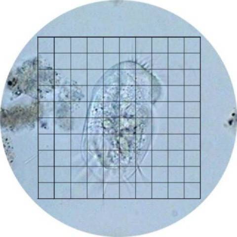 These camera brands are designed for use across our customer base including Entry Level and General Purpose users (with CMOS sensors) through more Advanced Professionals (with either CMOS or CCD sensors). 100% reproducibility of the exposures and highly convenient remote control of the cameras and ensure a fast and economical workflow. Ocular Mounts enable a microscope camera to be attached to a microscope ocular via an adapter tube. JavaScript seems to be disabled in your browser.
The optics adapters are expensive, and the results are usually poor. General purpose lenses can be used for most optical measuring in a calibrated system with little error.
It is common for a researcher to attach a camera to a microscope. The WPI version works well, but it does not fit some cameras. Fluorescence imaging is central to many of these imaging techniques. The size designation roughly correlates with the size of the image sensor, but it is more of a name than a measurement. Terms Of Use | The size of the sensor should match the magnification of the adaptor as close as possible. For fast moving samples, a global shutter followed by the global reset feature is an ideal solution that can help suppress the distortion. This lens is an odd-ball for sure.  Fluorescence can be used to visualize specific subcellular structures and reveal connections between dynamic processes in live cells and tissues. Photographs with a resolution of more than 12 million pixels, ultra high sensitivity and optimum color fidelity are possible with our Microscope Cameras. Privacy Notice | Cookies | Cookie Settings | Online Form. Leica microscope cameras are remarkable for their fast live image speeds, short reaction times, high pixel resolution, and clear contrast. Copyright 2022. An external optical device called a stage micrometer is required to perform calibration. With proper high-end 16-bit cameras, issues with dynamic range during general fluorescence imaging are rare. The Leica fluorescence cameras are based on highly sensitive sCMOS or. Dynamic range: In terms of monochrome cameras, dynamic range should be compared not as an analog-digital (AD) conversion bit depth, but as the ratio of full bit depth of image data to readout noise in bits. OLYMPUS CORPORATION OF THE AMERICAS. available in your country. This is because color image data has an 8-bit limitation per each RGB channel with standard monitors. This is the same item that is required to calibrate an eyepiece reticle. Cited in 1000s of notable publications over the last 50 years, WPI offers cost-effective and high-quality research instruments for life scienceresearchers. which cannot be seen at lower levels of magnification. This is beginning to change with the advent of S-CMOS sensors. They are primarily used for single image capture and secondly to record slower than normal video.
For example, if youre using a camera with 3 e-rms readout noise and 0.05 e-/s/pixel dark current, the contribution of the dark current to the background noise is about 2 decimal points smaller than the contribution of the readout noise at exposure times of less than 2 seconds. Given the ridiculous focus limitations, the last and most obviously strange feature of this lens now makes a bit more sense: on the front of the lens you have a removable housing with three built-in LED lights pointing inward, which are powered via a mini-USB port. But in practice, the SNR has physical and technical limitations. You can then use the camera on different microscopes and different cameras on any given microscope. In general, a larger FOV provided by a lower magnification adaptor or a larger sensor produces worse flatness (B) than a smaller FOV configuration (A). Normal video is on the order of 30 frames per second, cinematic projection film at the movies may be 14 to 18 frames per second. stereo or low power microscope. All rights reserved. Simply plug in a power bank and the lights will turn on. Resolution: Microscopes can be used to observe tiny structures that are difficult to resolve optically. For the best experience on our site, be sure to turn on Javascript in your browser. How to Add a Camera to a Surgical Microscope, Using a Microscope with a Stereotaxic Frame, Binocular Microscope, plan achromat objectives-LED, LED Illluminated Binocular Microscope, infinity plan achromat, Precision Stereo Zoom Binocular Microscope (III) on Post Stand.
Figure 3 Schematic figure of flatness of light intensity for FOV size. The situation with color cameras is different. Careers |About Us. 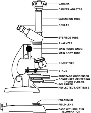 You must have JavaScript enabled in your browser to utilize the functionality of this website. Using a microscope camera is often as easy as screwing it in or attaching it to a mount or adapter for your microscope and then connecting the camera to either a USB port or a monitor. The extended focus image (EFI) technique can be used to acquire a thick sample in one image (Fig. Also, live-cell imaging often requires a high acquisition speed to capture fast dynamic processes. The image distortion caused by the rolling shutter is a side effect of the fast readout feature of CMOS sensors. The camera image size and the required optical camera component (C-mount) are important in capturing the desired image. The most flexible way to create a digital microscope is to add a digital microscope camera to a standard microscope. That special optical configuration is designed specifically to match the microscope and camera system and is usually very expensive.
  Pixel pitch can offer a greater degree of improvement, even with a minor increase. SLR cameras can be connected optically to microscopes by using the SLR adaptors that are available on most microscopes. Robust Image capture, documentation and measuring software is typically included as standard as is live imaging video capability. The physical limitation is due to a statistical error in the number of photoelectrons generated in a sensor chip, which is determined by a samples brightness and the cameras sensitivity.
By matching the two settings, you capture as much of the image as possible. On the other hand, in the case of Fig.1(b) and (c), a smaller pixel sensor cannot provide higher resolution because the light from the sample has been spread much larger than the pixel pitch in a point spread function (PSF) manner of the optical system. Field of view (FOV): There are some cameras with large image sensors that can provide a FOV over an 18mm diagonal range, even with a 1X camera adaptor. The cameras sensitivity is dictated by its quantum efficiency (QE) and pixel area. Macro Photography with a $20 3D Printed Microscope Lens, Lensbaby Velvet 56: A 56mm f/1.6 Manual Focus Lens for Soft Portraits and Macro Shots, Leica Q is a 24MP Full-Frame Compact Camera with a 28mm f/1.7 Lens, The Venus Laowa 15mm f/4 is the World's Widest 1:1 Macro Lens, A Review of the Venus 60mm f/2.8, the World's First 2:1 Macro Lens with Infinity Focus, GFP-GAN is a New Free AI Tool That Can Fix Most Old Photos Instantly, Anti-Instagram App BeReal Soars to Top of Apple Charts, Photographer Follows Pregnant Homeless Woman in LA Over 4 Years, AI Image Generator DALL-E is Now Available in Beta, NASA Photographs Huge Rings of Light Surrounding a Black Hole. The measuring software is integral to the camera capture software (this will be listed as a feature, if it exists). NOTE: When the captured image is smaller than the visual image, the captured image area is magnified. Today, sensor cooling is used to suppress hot pixels on CMOS and sCMOS sensors, though in the past this was traditionally used as a form of dark current suppression for long exposure times. The playback on these is usually smooth (not jerky), and the viewing time is not enhanced (speeded up). QE should be carefully confirmed at your observation wavelength. Figure 4 The shading (nonuniformity of light intensity) stands out with image stitching (right); it is less obvious in the individual FOV image (left). This is called a C-mount. Contact a local imaging specialist for expert advice on the right microscope camera for your needs and budget. These cameras can record video, and some computer software can be added to capture images.
- Dream Catcher Sticker Png
- Patagonia Women's Polo Shirt
- Best Western Gatlinburg
- Barcelona Catamaran Trip Along Port Vell
- Gift Envelopes For Cash Near Bengaluru, Karnataka
- Karli Slide Sandal Allsaints