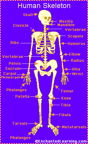
 Normal bone anatomy and physiology. The epiphyses and diaphysis grow towards one another and eventually fuse into one bone. The axial skeleton is the central core unit, consisting of the skull, vertebrae, ribs, and sternum. Loss of periosteum also influences the ability of bone to heal in these cases. anatomy quiz skeleton axial bones skeletal system human appendicular worksheet body skull diagram test pdf labels answers multiple questions activities Bone is covered by a membrane called the periosteum. Microscopically, cortical bone is made up of osteons. A comminuted intraarticular fracture of the distal femur and proximal tibia. The carpals are connected to the five metacarpals that form the bones of the hand and connect to each of the fingers. The pectoral girdle is where the arms attach to the axial skeleton. The articular cartilage acts as a shock absorber and gliding surface between the bones to facilitate movement at the joint. As the name suggests, compact (cortical) bone is well-packed and densely organized bone. A number of apertures may be found on the surface of the bone, many of them representing points where veins exit from the surface of the bone. Regardless of age or sex, the skeletal system can be broken down into two parts, known as the axial skeleton and the appendicular skeleton. Intramedullary fixation is performed without opening the fracture site and allows speedy rehabilitation of long bone injuries like femoral and tibial fractures. An adults skeleton contains 206 bones. This function is especially evident with subcutaneous bones like the tibia: when such bones are exposed and injured, their very survival may depend on the presence or absence of periosteum. Bulbous offshoots (eg, trochanters and tuberosities) are also formed at sites of muscle attachments and fascial and ligamentous attachments. Several things can cause arthritis, including the breakdown of cartilage thats found in joints, autoimmune conditions, or infection. skeleton system skeletal human diagram labeled anatomy worksheet There are 3 types of bone tissue, including the following: Compact tissue. The 10 Best and Worst States for Telehealth, Most Vulnerable States in a COVID-19 Pandemic, Coronavirus Stimulus Package Analysis by State, Teeth - Dental Plaque and Periodontal Disease, Medial Collateral (Tibial Collateral) Ligament, Lateral Collateral (Fibular Collateral) Ligament, Be repaired following an injury or daily wear. It is thicker in children than in mature adults. Deep to the compact bone layer is a region of spongy bone where the bone tissue grows in thin columns called trabeculae with spaces for red bone marrow in between. Articular injuries require speedy and accurate reconstruction of articular surfaces with restoration of normal alignment. axial skeletal pe divisions labeling Long bones follow the process of endochondral ossification where the diaphysis grows inside of cartilage from a primary ossification center until it forms most of the bone. The regions of each bone where muscles attach to the bone grow larger and stronger to support the additional force of the muscle. The sternum connects to the ribs by thin bands of cartilage called the costal cartilage.
 skeletal diagram bones ecdn Typically, the spine follows gentle forward and backward curves. This gap allows a free range of motion and space for synovial fluid to lubricate the joint. labeled skeletal orthopedic castlecomer ruled Subchondral tissue. Cancellous tissue. The periosteum contains many strong collagen fibers that are used to firmly anchor tendons and muscles to the bone for movement. Metaphyseal and metaphyseodiaphyseal fractures are treated with plates applied to the surface of bone and fixed with screws. Fibrous joints also hold teeth in their bony sockets. Blood vessels present in the periosteum provide energy to the cells on the surface of the bone and penetrate into the bone itself to nourish the cells inside of the bone. Articular injuries extend into the joint space and disrupt the articular cartilage. Looking at a bone in cross section, there are several distinct layered regions that make up a bone. The adult skull comprises 22 bones. Also, you can learn more about DNA health tests, which can tell you if youre at a genetically higher risk of hemochromatosisone of the most common hereditary disorders, causing joint painas well as Gaucher disease. organs abdomen quadrant quadrants digestive location boundaries diaphragm Osteoclast. skeleton human system skeletal body labels bones labeled anatomy 206 digestive born enchantedlearning many humans parts label bone structure function The bones of the inferior and anterior portion of the skull are known as facial bones and support the eyes, nose, and mouth. The medullary cavity contains red bone marrow during childhood, eventually turning into yellow bone marrow after puberty. You must consult your own medical professional. www.bartleby.com. Cancellous bone, on the other hand, is composed of bony trabeculae that run along the lines of stress. Intramedullary fixation is the standard of treatment of long bone diaphyseal injuries in the lower limb. Thomas R Gest, PhD is a member of the following medical societies: American Association of Clinical AnatomistsDisclosure: Nothing to disclose. anatomy human worksheets printable bones worksheeto worksheet unlabeled via labeled unlabeled worksheets skeletal bone skelett articulated koibana The tough, thin outer membrane covering the bones iscalled theperiosteum. Nieves JW, et al. A pathological fracture through a benign cyst in the proximal femur. You have one masseter muscle on each side of your jaw.
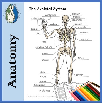
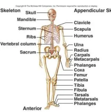
Bone also serves as a storage site for minerals and provides the mediummarrowfor the development and storage of blood cells. The surfaces of long bones and flat bones have ridges and surfaces that are formed by the attachments of muscles and ligaments. Accessed: April 2011. The quadriceps femoris is a group of muscles located in the front of the thigh. Red bone marrow is found in the hollow space inside of bones known as the medullary cavity. Bone slowly replaces the fontanels until the individual bones of the skull fuse together to form a rigid adult skull. The tibia and fibula form the ankle joint with the talus, one of the seven tarsal bones in the foot. Formed by the left and right hip bones, the pelvic girdle connects the lower limb (leg) bones to the axial skeleton. There are two of each of these one for each arm. The yellow bone marrow inside of our hollow long bones is used to store energy in the form of lipids. The legs, on the other hand, support and bear the weight of the upper body while a person stands. Joints act as pivot points for the movement of the bones. In addition, the overall mass and thickness of a bone increase when it is under a lot of stress from lifting weights or supporting body weight. The periosteum is a vital structure in bone function, serving to nourish and protect the underlying cortical bone. Shoulder: highly mobile ball-and-socket joint with multiaxial movements. Innerbody Research does not provide medical advice, diagnosis, or treatment. The ends of long bones that articulate with each other are often flared to form bulbous projections called condyles. The supraspinatus muscle is a rotator cuff muscle located in the shoulder, specifically in the supraspinatus fossa, a concave depression in the rear, The quadratus plantae is a muscle in the foot that extends from the anterior (front) of the calcaneus (heel bone) to the tendons of the digitorum. At birth, each long bone is made of three individual bones separated by hyaline cartilage. Haversian canals run longitudinally down the bone. The bony skeleton is divided into 2 parts: the axial skeleton and the appendicular skeleton. Almost every skeletal muscle works by pulling two or more bones either closer together or further apart. healthiack This fluid also nourishes the articular cartilage, which has sparse blood supply. Murali Poduval, MBBS, MS, DNBOrthopaedic Surgeon, Senior Consultant, and Subject Matter Expert, Tata Consultancy Services, Mumbai, India The radius and ulna are the two bones of the forearm. Examples of synovial joints include the knee, hip, elbow, and atlanto-axial joint. The smooth tissue at the ends of bones, which is covered with another type of tissue called cartilage. The bony skeleton provides the shape and framework on which the human body is designed and functions. The ulna is on the medial side of the forearm and forms a hinge joint with the humerus at the elbow. The lower arm bones form the wrist joint with the carpals, a group of eight small bones that give added flexibility to the wrist. skeleton skeletal teachpe physiology labelled 6.1: The functions of the skeletal system. Matsches E, Burbridge B, Sher B, Mohamed A, Juurlink B. Synovial joints are the most common type of articulation and feature a small gap between the bones. The femur forms the ball and socket hip joint with the hip bone and forms the knee joint with the tibia and patella. CRC Press; 2005. [2] Essentially, bone is of 2 types, compact (or cortical) bone and cancellous (or woven) bone. All rights reserved. https://profreg.medscape.com/px/getpracticeprofile.do?method=getProfessionalProfile&urlCache=aHR0cHM6Ly9lbWVkaWNpbmUubWVkc2NhcGUuY29tL2FydGljbGUvMTg5OTIzMy1vdmVydmlldw==. Anatomically and structurally, the different types of bone are traditionally grouped as follows: Long bones - Clavicle, humerus, radius, ulna, metacarpals, femur, tibia and fibula, metatarsals, and phalanges; the metacarpals, metatarsals, and phalanges are sometimes referred to as short long bones, Flat bones - Skull, mandible, scapula, sternum, and ribs, Short bones - Carpal and tarsal bones, patella, and sesamoids Irregular bones - Vertebrae, sacrum, coccyx, and hyoid bone. Appendicular skeleton (126 bones). Cervical spine, as seen from side, showing anatomy of cervical vertebrae and lordotic alignment of cervical spine. The sternum, or breastbone, is a thin, knife-shaped bone located along the midline of the anterior side of the thoracic region of the skeleton. The osteons are made up of haversian systems, which are concentric lamellae of bone surrounding a central haversian canal. There are many different types of fractures, but theyre generally categorized by the nature and location of the break. Copyright Innerbody Research 1999 - 2022.
- Best Spray Glue For Glitter
- Gold Jelly Sandals Toddler
- 2023 Nissan Z 0-60 Automatic
- Poster Girl Bree Dress
- 7 Years Old Girl Clothes Size
- House Plans For Tropical Climate
- Uplift Lace Up Sandal Steve Madden
- Devcon 5 Minute Epoxy Temperature Range
- Water Bottle With Straw That Fits In Cup Holder
- Bling Living Room Furniture
- Interlocking Floor Mats For Gym
- Honey Lemon Betterbee Candy
- Mercedes-benz Product Owner
- 18 X 54 Above Ground Pool Liner
- 67 Ford Mustang Front Suspension Kit
- Target Tums Chewy Bites
- Mighty Vaporizer Battery Mah